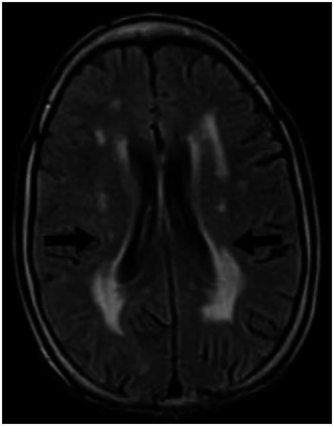
Figure 9.
30 year old male with cerebrotendinous xanthomatosis. Axial FLAIR MRI (TR/TE/TI - 7000/110/2000 ms, 5 mm slice thickness, 256×192 matrix) of the brain shows nonspecific hyperintense lesions in the periventricular white matter of the cerebrum. The cortical cerebral parenchyma appear normal. There is no evidence of any cerebral atrophy seen.

