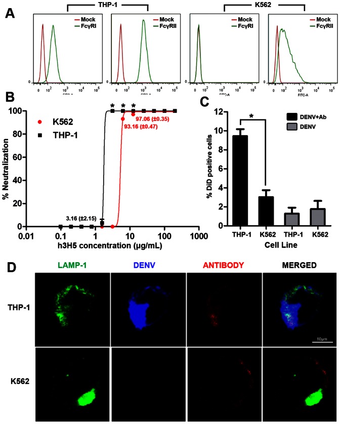
Figure 1. Absence of FcγRI engagement is associated with increased antibody requirement for DENV neutralization.
(A) Flow cytometry data of both FcγRI and FcγRII (green histogram) in THP-1 and K562. Mock (red histogram) represents staining with secondary antibody only. (B) Neutralization profile of DENV using various concentrations of h3H5 antibody in THP-1 (Black) and K562 (Red) at 72 h post infection, quantified by plaque assay. Unless indicated, the mean value of neutralization is either 0% or 100%. (C) Percentage of internalized DiD-DENV (% DiD positive cells) in complex with h3H5 antibody is represented by black bar at concentrations that mediated complete neutralization in THP-1 (3.125 µg/mL) and K562 (25 µg/mL), DiD-DENV without antibody is represented in grey bar, as assessed by confocal microscopy at 30 mins post infection. (D) Subcellular localization of DiD-DENV opsonized at h3H5 antibody concentrations required for complete neutralization in THP-1(3.125 µg/mL) and K562 (25 µg/mL). LAMP-1 is in green, DiD-DENV is in blue and h3H5 antibody is in red. Scale bar is 10 µm. Data are represented as mean ± s.e.m. * p<0.01. Results presented are mean of three independent experiments, each with biological triplicates.

