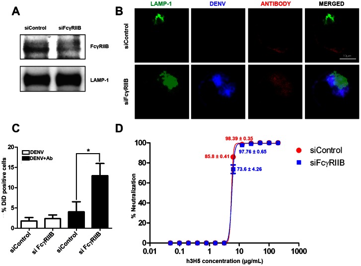
Figure 2. FcγRIIB knockdown did not result in additional increase in antibody requirement for DENV neutralization.
(A) K562 transfected with either control siRNA or FcγRIIB siRNA. The reduction in FcγRIIB expression was detected in cell lysate by western blot. LAMP-1 served as a loading control. (B) Subcellular localization of neutralized DENV immune complexes in K562 treated with either control siRNA or FcγRIIB siRNA at 30 mins post infection. LAMP-1 is in green, DiD-DENV is in blue and h3H5 antibody is in red. Scale bar is 10 µm. (C) Percentage of DiD positive cells in K562 treated with either control siRNA or FcγRIIB siRNA with DiD-DENV using 25 µg/mL antibody (black bar) or without antibody (white bar) after 30 mins post infection, assessed by confocal microscopy (D) Neutralization profile of h3H5 against DENV in K562 treated with either siRNA control (red) or siRNA against FcγRIIB (blue) at 72 h post infection, assessed by plaque assay. Unless stated, the mean percent neutralization is either 0% or 100%. Data are represented as mean ± s.e.m. * p<0.01. Results presented are mean of three independent experiments, each with biological triplicates.

