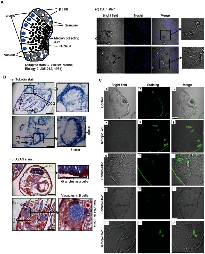
Figure 4. Differential localizations of Bamcp20k-1 and Bamcp20k-2 in cyprid cement glands.
(A) The diagram shows the composition of a cement gland in Balanus balanoides. Two types of cement cells (α and β) were shown in the cement glands. (B) Cyprids were fixed in 4% PFA and then sectioned after embedding in wax. A section of 4 µm from Amphibalanus Amphitrite was shown. (a) Toluidin stain, Scale bar: 50 µm; (b) AZAN stain, Scale bar: 50 µm; (c) DAPI stain. Nuclei were stained with DAPI. Scale bar: 10 µm. CE: compound eye; CG: cement gland; GN: granules; NC: nuclei. (C) Bamcp20k-1 and Bamcp20k-2 were localized at different cement granules. Sections were immunostained with antibodies against Bamcp20k-1 and Bamcp20k-2. For (a) to (c), the sections stained only with secondary antibody were used as negative controls; for (d) to (i), the sections were stained with an antibody against Bamcp20k-1; for (j) to (o), the sections were stained with an antibody against Bamcp20k-2. Scale bar: 30 µm.

