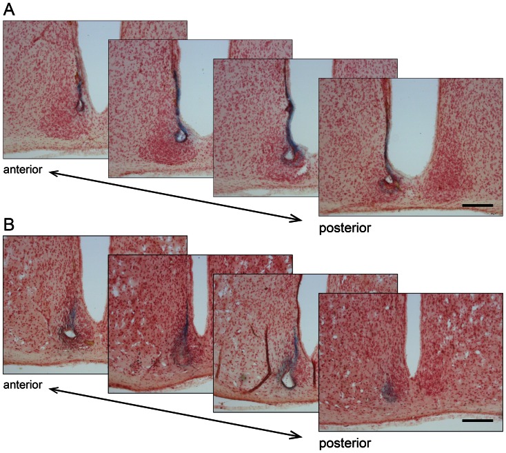
Figure 1. Histological examination of recording sites in the ventral and dorsal SCN.
Representative serial coronal sections (25 µm, every other section shown) of a wild-type (A) and Per1−/− (B) SCN following MUA recording. Sections were stained with neutral-red and blue spots of deposited iron are the recording sites. Recording sites were present in the ventral and dorsal SCN. The anterior to posterior orientation of the sections is indicated. Scale bar represents 200 µm. The data in A and B correspond to the data shown in Fig. 2A and B, respectively.

