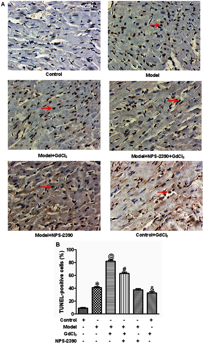
Figure 4. Representative illustration of TUNEL staining in cardiomyocytes apoptosis.
(A) Nuclei with brown staining were TUNEL-positive cell, which was defined as apoptotic cell. Magnification at 400 ×. (B) Statistical analysis of cardiomyocytes apoptosis (n = 8). *P<0.05 compared with control group; @P<0.05 compared with model group; #P<0.05 compared with model+GdCl3 group; &P<0.05 compared with control group.

