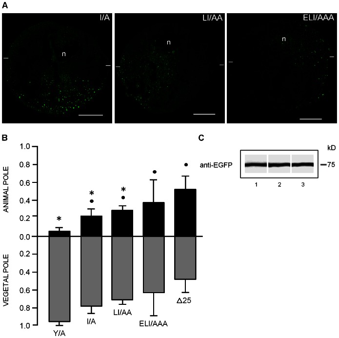
Figure 8. Retention and polarization of GIRK5 mutants bearing the I/A mutation.
A) Confocal microscopy assays and B) Quantification of fluorescence show that removal I22 residue in GIRK5, as shown in I/A and LI/AA, causes loss of polarization, whereas the ELI/AAA variant is targeted to both poles like Δ25. Scale bar: 250 µm. Error bars correspond to mean ± SD, n = 4–6. A circle and an asterisk indicate significant differences compared to oocytes expressing Y/A and Δ25, respectively (P<0.05; One-Way ANOVA). The statistic significance between samples is the same for the animal and vegetal pole. C) Immunoblot analysis of EGFP-GIRK5 mutants with bands corresponding to the expected weight of 75 kDa: 1) I/A, 2) LI/AA, 3) ELI/AAA.

