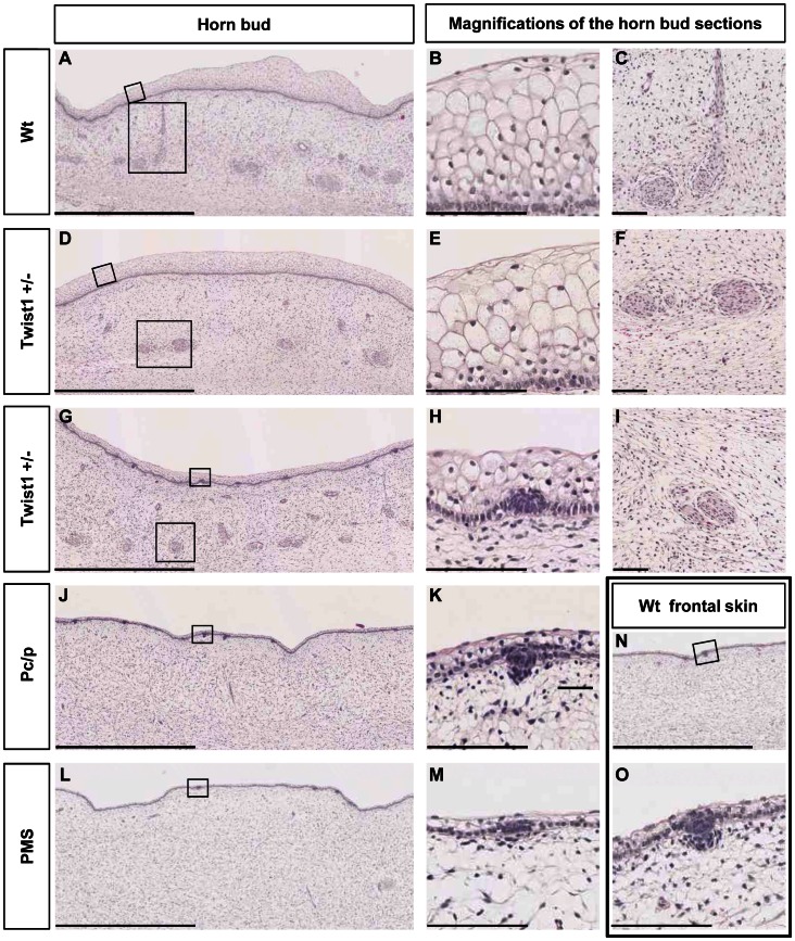
Figure 4. Histological analyses of horn buds and forehead skin from wild-type fetuses and fetuses affected by different horn-defect syndromes.
(A), (D), (G), (J) and (L) Histological sections of horn buds of a wt, two TWIST1+/−, a PC/p and a PMS fetus, respectively. (B) and (C), (E) and (F), and (H) and (I) Magnifications (X10 and X3, respectively) of (A), (D) and (G). (K) and (M) Magnifications (X10) of (J) and (L). (N) Histological section of the forehead skin of a wt fetus. (O) Magnification (X10) of (N). Scale bars in (A), (D), (G), (J), (L) and (N) represent 1 mm, whereas scale bars in (B), (C), (E), (F), (H), (I), (K), (M) and (O) represent 100 µm.

