
Figure 3. Immunodetection of FAR proteins in Meloidogyne incognita pre-parasitic J2 and during parasitism of Arabidopsis thaliana roots.
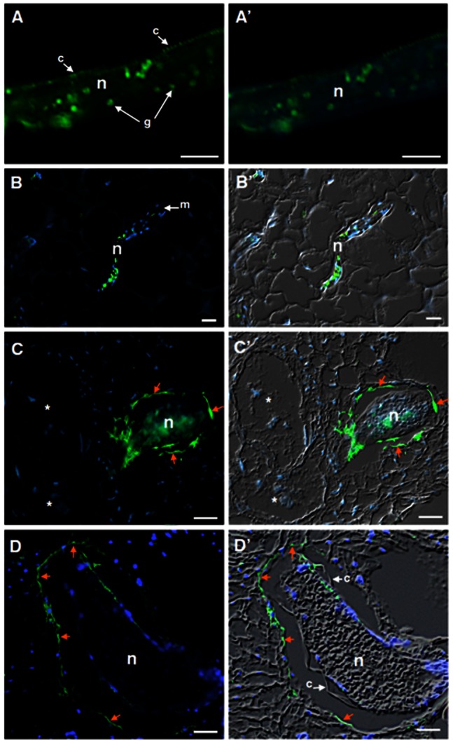
(A) Cross nematode sections of pre-parasitic J2 displaying the protein localization at the nematode cuticle surface and circular granules structures within the posterior nematode body. (B) Localization of FAR proteins during nematode migration (B-B′), and nematode sedentary stages at 10 DAI (C-C′) and 21 DAI (D-D′) within the roots of A. thaliana. Arrows point out the accumulation of FAR along the nematode cuticle and adjacent cells surrounding the nematode body at 10 and 21 DAI. Micrographs on the left are overlays of Alexa-488 fluorescence (green) and DAPI-stained nuclei (blue). Micrographs on the right are overlays of an Alexa-488 fluorescence (green), DAPI-stained nuclei (blue) and differential interference contrast (grey). c, cuticle, g, granules, n, nematode, m, metacorpus, * giant cell. Bars = 10 µm.