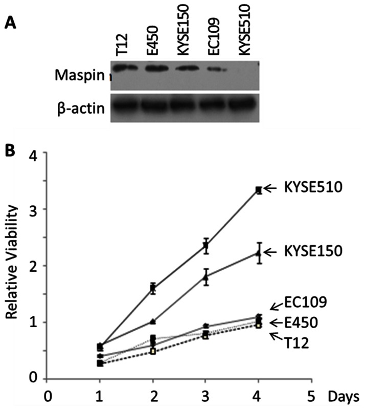
Figure 3. The correlation of maspin expression in established human ESCC cell lines with lower rates of proliferation in vitro.

(A) Western blotting of maspin in the indicated ESCC cell lines. Twenty-five micrograms of total lysate protein were loaded in each lane. Western blotting of the same membrane for house-keeping β-actin was used to assess the loading variation. (B) MTT assay of the proliferation of ESCC cell lines. The data at each time point represent the average of three independent repeats. The error bars represent the standard deviation.
