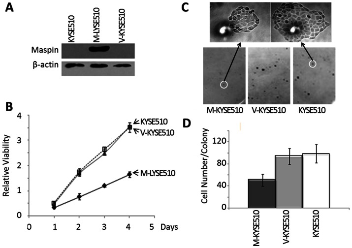
Figure 4. Characterization of stably transfected KYSE510 cell lines.
(A) Western blotting of maspin and housekeeping protein β-actin in the total lysates of parental KYSE510, M-KYSE510, and V-KYSE510 cells. (B) MTT assay of the proliferation of parental KYSE510, M-KYSE510, and V-KYSE510 cells, cultured in the maintenance media. (C) Representative staining of single cell-derived colonies (bottom) and the magnified image of the highlighted colonies (top) from the colony formation assay. (D) Quantification of colonies with more than >100 cells/colony based on counting under microscope in the colony formation assay. Data represent the average of three independent repeats. Error bars represent the standard deviation. The difference between M-KYSE51 and V-KYSE510 (or parental KYSE510) was statistically significant (p<0.001).

