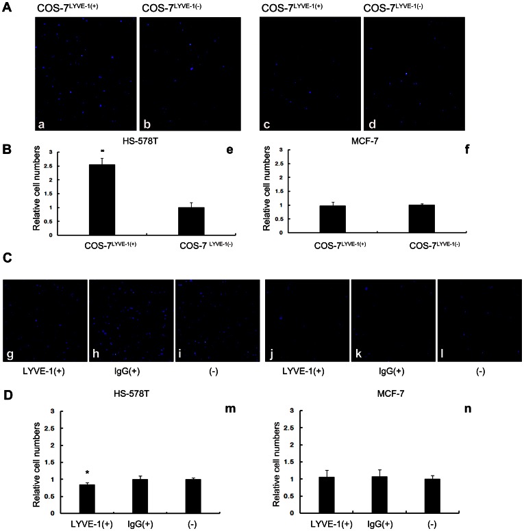
Figure 2. Breast tumor cells expressing high or low levels of HA adhere differently to cells under static conditions.
(A) Adhered HS-578T and MCF-7 cells appear as blue spheres (a–d). DAPI-labeled HS-578T cells were more adherent to the confluent monolayer of COS-7LYVE-1(+) cells compared with COS-7LYVE-1(−). MCF-7 cells adhered similarly with both COS-7LYVE-1(+) and COS-7LYVE-1(−) cells. (B) Quantitative evaluations of HS-578T and MCF-7 cell adhesion on the monolayer of COS-7 cells are shown, and each assay was performed in triplicate. The results are shown as the mean ± SD of triplicate wells from a typical experiment and are expressed as a fold increase relative to COS-7 cells transfected with the control vector pEGFP-N1. *P<0.05 versus indicates transfected pEGFP-N1 vector control COS-7 cells. (C) Adhered HS-578T and MCF-7 cells appear as blue spheres (g–l). DAPI-labeled HS-578T cells were adherent to the confluent monolayer of SVEC4-10 cells before and after blocking with LYVE-1 antibody, isotype antibody. (D) Quantitative evaluations of the data from HS-578T and MCF-7 cell adhesion on the monolayer of SVEC4-10 cells are shown, and each assay was performed in triplicate. The results are shown as the mean ± SD of triplicate wells from a typical experiment and are expressed as a fold decrease relative to SVEC4-10 cells without treatment. *P<0.05 versus SVEC4-10 cells without treatment.

