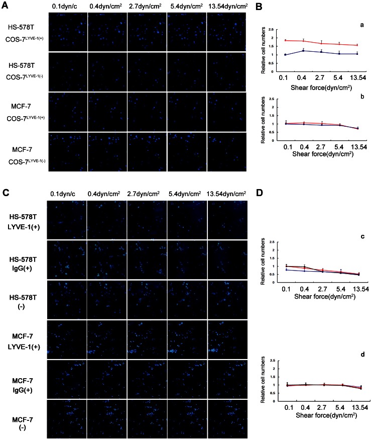
Figure 4. HA-independent adhesion of HS-578T and MCF-7 cells was visualized under flow conditions.
HS-578T and MCF-7 cells were perfused over COS-7LYVE-1(+) and COS-7LYVE-1(−) cells at wall shear stresses of 0.1, 0.4, 2.7, 5.4 or 13.54 dyn/cm2 for 2 min. (A) The number of adherent cells in the same field is shown as blue spheres. (B) The results are shown as the relative cell numbers of HS-578T cells (a) and MCF-7 cells (b) captured by COS-7LYVE-1(+) (red line) to COS-7LYVE-1(−) (blue line) cells. HS-578T and MCF-7 cells were perfused over a confluent monolayer of SVEC4-10 cells before and after blocking with LYVE-1 antibody, isotype antibody at wall shear stresses of 0.1, 0.4, 2.7, 5.4 or 13.54 dyn/cm2 for 2 min. (C) The number of adherent cells in the same field is shown as blue spheres. (D) The results are shown as the relative cell numbers of HS-578T cells (c) and MCF-7 cells (d) captured by SVEC4-10 cells blocking with LYVE-1 (blue line), SVEC4-10 cells blocking with isotype control (red line) to SVEC4-10 cells without treatment (black line). The error bars represent the mean ± SD of the number of cells bound. Data are representative of 3 independent experiments.

