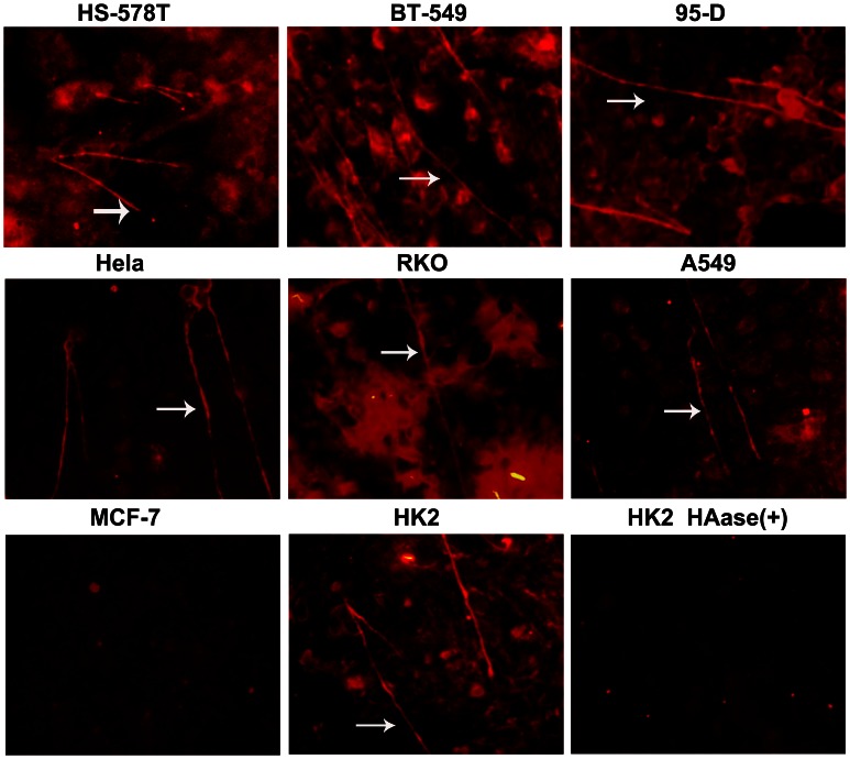
Figure 5. The tumor cell surface HA content is rich, and HA forms cable structures.
Confluent monolayer cells were grown in the presence of 10% (v/v) FCS before fixation with methanol and the detection of HA by the addition of bHABP. The sections were imaged by inverted fluorescence microscopy (×20 objective). HA cables are highlighted with white arrows. Original magnification ×20. Streptomyces hyaluronidase was added to the cells before fixation. The HA cables were degraded without detection.

