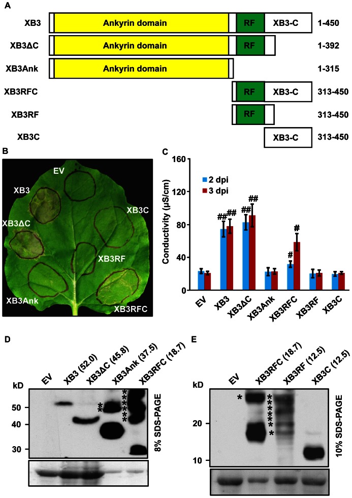
Figure 3. The RING finger (RF) motif of XB3 is required for its cell death activity.
(A) Schematic diagram showing structure of XB3 and its truncation mutants used for agroinfiltration. Domains were as described by [14]. Numbers on the left indicate amino acid positions of each product in the full-length XB3. A 3xFLAG epitope tag was individually fused to the C-terminus of each protein. (B) Phenotypes induced by the expression of XB3 or its truncation mutants in N. benthamiana leaves. (C) Quantification of the cell death induced by the indicated proteins. Agroinfiltration and electrolyte leakage assays were performed as described in the Figure 2 legend. Data sets with pound signs indicate statistically significant differences from the control (EV) as calculated by Student's t test (#: P<0.05; ##: P<0.01). (D, E) Protein blot analyses showing the levels of XB3 and its truncation mutants in the infiltrated leaves. Total protein extracts were prepared 40 hours after infiltration and immunoblotted with anti-FLAG M2 antibody (Top). The same blot was stained with Ponceau S to show sample loading (Bottom). The theoretical molecular weights (kDa) of each protein are shown in parentheses. Percentages of the SDS-PAGE gels used for resolving the proteins are indicated. Asterisks denote high molecular weight products derived from the indicated mutants.

