FIG. 2.
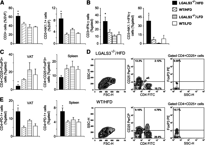
Increased type 1 T and NKT cells and reduced Tregs in VAT of HFD-fed LGALS3−/− mice. A: Increased percentages of CD3+ T cells and in VAT of LGALS3−/− mice fed HFD compared with other groups after 11 weeks. CD3+NK1.1+ NKT cells in VAT of LGALS3−/− mice fed HFD were significantly increased compared with WT mice on both diet conditions. B: LGALS3−/− mice fed HFD have significantly increased frequencies of CD3+IFN-γ+ in VAT compared with LFD-fed mice of both genotypes, whereas CD3+NK1.1+ NKT cells that express IFN-γ were significantly higher compared with other experimental groups. C: Adipose tissue regulatory CD4+CD25+FoxP3+ T cells are reduced, and the trend toward decreased Tregs is observed in spleens (P = 0.071) of LGALS3−/− vs. WT mice fed HFD. D: Representative fluorescence-activated cell sorter plot of CD4+CD25+FoxP3+ T cells in VAT from HFD-fed LGALS3−/− and WT mice. E: Significantly increased percentages of CD4+PD-1+ cells in VAT and spleen from HFD-fed LGALS3−/− mice vs. WT mice on both diet conditions. The results are shown as the means ± SEM for four to six animals per group. FSC-H, forward-scattered light; FITC, fluorescein isothiocyanate; SSC-H, side-scattered light in flow cytometry; SVF, stromal vascular fraction. *P < 0.05.

