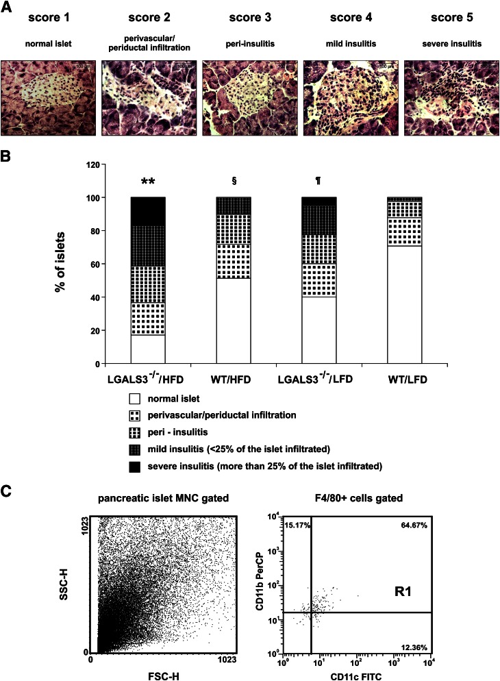
FIG. 5.
Histological analysis of infiltrating mononuclear cells in pancreatic islets. A: Hematoxylin-eosin staining was performed on pancreatic tissue sections. Representative images of insulitis scores in LGALS3−/− mice fed HFD for 11 weeks. Original magnification ×40. Scale bars, 100 µm. B: Pancreatic islet inflammation (insulitis) was graded from 1 to 5, according to the extent of peri- and intraislet infiltration by mononuclear leukocytes as follows: 1, no islet infiltration; 2, peri-vascular/periductal islet infiltration; 3, peri-insulitis; 4, mild insulitis (<25% islet area infiltrated); 5, severe insulitis (>25% islet area infiltrated). LGALS3−/− mice on HFD had a significantly increased percentage of islets with severe insulitis compared with other experimental groups. LGALS3−/− mice on LFD had a significantly higher percentage of severe insulitis compared with WT mice on both diet conditions. The results are shown as percentages of islets with insulitis derived from four to six mice per group. **P < 0.001; ¶P < 0.05; §P < 0.05. C: FACS plots of mononuclear cells isolated from the pooled pancreata (n = 5) of LGALS3−/− mice fed HFD. The dot plots depict forward-scattered light (FSC) and side-scattered light in flow cytometry (SSC) (left) and the CD11b+CD11c+ cells among gated F4/80+ cells (right). In FACS analyses, mononuclear infiltrates were not found in mice from other experimental groups. (A high-quality color representation of this figure is available in the online issue.)

