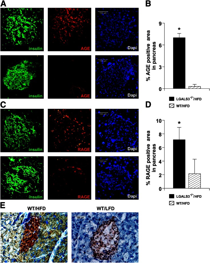
FIG. 7.
Increased expression of AGE and RAGE in pancreatic islets of HFD-fed LGALS3−/− mice. A: Immunofluorescence staining for insulin (green) and AGE (red) together with DNA staining with DAPI (blue) in pancreatic islets from representative LGALS3−/− (top) and WT mice fed an HFD (bottom). B: Evaluation of % AGE-positive areas was performed on IHC-stained tissue sections from four to six mice per group. C: Immunofluorescence staining for insulin (green) and RAGE (red) together with DNA staining with DAPI (blue) in pancreatic islets from representative LGALS3−/− (top) and WT mice fed an HFD (bottom). D: Evaluation of % RAGE-positive areas was performed on IHC-stained tissue sections from four to six mice per group. E: Representative IHC image of Gal-3 expression in HFD- or LFD-fed WT mice. The results are representative of two to three repeated experiments. *P < 0.05.

