FIG. 2.
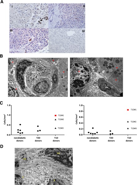
A: Neutrophils are detected by immunoperoxidase in the exocrine pancreas. Representative neutrophil stainings of sections from donor no. 1 with T1D (I and II), donor no. 3 with T1D (III), and a nondiabetic donor (IV) are shown. These pancreas-infiltrating neutrophils localize mainly at the level of very small blood vessels (panels I and III) and, to a lesser extent, adjacent to acinar cells (panel II). B: Representative sections of the pancreas collected from donor no. 1 with T1D, analyzed using electron microscopy, and showing a granulocyte (GN) and a lymphocyte (L) in a microvessel of the exocrine tissue (left panel), and a GN adjacent to acinar cells (right panel). In both panels, red stars identify pancreatic acinar cells. C: The frequency of pancreatic neutrophils is determined by immunoperoxidase on pancreatic paraffin sections. The numbers of myeloperoxidase positive cells per square millimeter in the pancreas (left panel) and within the small blood vessels (right panel) are shown. At least 20 fields per donor have been examined. D: A representative section of the pancreas collected from donor no. 1 with T1D, analyzed using electron microscopy and showing GNs adjacent to β-cells (β).

