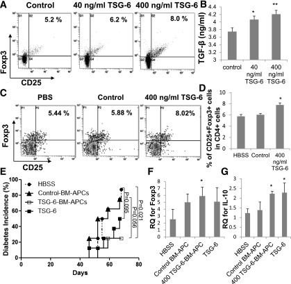
FIG. 6.
TSG-6-BM APCs delayed diabetes in an adoptive transfer model. Treg flow analysis (A) and TGF-β expression (B) 4 days after TSG-6-BM APC (400 ng/mL, 5 × 10⁵ cells/mL) and CD4+ T-cell (106 cells/mL) cocultures. Splenic Treg representative flow cytometry analysis (C) and quantification (D) 5 d after vehicle control (PBS, n = 4), control-BM APCs (control, 1 × 10⁶, n = 3), or TSG-6-BM APCs (400 ng/mL, 1 × 10⁶, n = 3) were intravenously infused in 5-week-old female NOD mice. Values are means ± SD. *P < 0.05, **P < 0.005 by two-tailed Student t test. E: Diabetes incidence after diabetogenic splenocytes (107 cells/mouse) pooled from 11-week-old female NOD mice that were cotransferred with control-BM APCs (106 cells/mouse, n = 8), TSG-6-BM APCs (106 cells/mouse, n = 8), TSG-6 (50 µg/mouse, n = 8), or vehicle control (HBSS, n = 8). TSG-6–treated animals received an additional intravenous infusion of TSG-6 (50 µg/mouse) 1 week posttransfer. HBSS vs. TSG-6-BM APCs, P = 0.021; control-BM APCs vs. TSG-6-BM APCs, P = 0.056; HBSS vs. TSG-6, P = 0.095 by Kaplan-Meier estimator. foxp3 (F) and il-10 (G) expression in splenocytes isolated from mice from E (n = 3–5) at 70 days. Values are means ± SD. *P < 0.05 by two-tailed Student t test. RQ, relative quantification.

