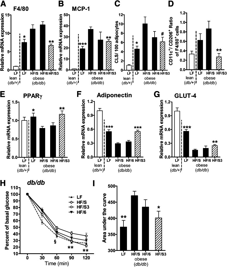
FIG. 4.
Dietary n-3 PUFA treatment attenuates adipose tissue inflammation and improves insulin sensitivity. Lean (db/+) mice were fed an LF diet and obese (db/db) animals were fed an LF diet or three different isocaloric HF diets: 1) HF/S diet rich in saturated/monounsaturated fatty acids; 2) HF/6 diet rich in n-6 PUFA; and 3) HF/S3 (HF/S diet supplemented with n-3 PUFA) for 6 weeks. Gonadal adipose tissue expression of the genes for macrophage marker F4/80 (Emr1) (A) and MCP-1 (Ccl2) (B) after dietary treatment was analyzed (n = 10 animals per group). (C) CLS formation, a hallmark of obesity-associated inflammation, was assessed by MAC-2 staining, and the number of CLS in gonadal adipose tissue was calculated per 100 adipocytes (n = 5 animals per group). (D) Flow cytometry analysis of the CD11c+CD206−-to-CD11c−CD206+ ratio of adipose tissue macrophages (F4/80+ cells) obtained from stromal vascular fractions of the db/+ LF control and db/db animals after dietary treatment with indicated diets (n = 8 animals per group). Expression of the genes for PPARγ (Pparg) (E), adiponectin (Adipoq) (F), and GLUT-4 (Slc2a4) (G) after dietary treatment in gonadal adipose tissue (n = 10 animals per group). (H and I) Insulin sensitivity was determined in db/db mice after LF control, HF/S, HF/6, or HF/S3 diet. Blood glucose was measured before and 30, 60, 90, and 120 min after intraperitoneal injection of insulin (2.0 units/kg body weight; n = 7–9 animals per group), and the area under the curve was calculated (n = 7–9 animals per group). For statistical analysis, db/db mice were compared with those fed the HF/S diet. All data are mean ± SEM. #P = 0.067; *P < 0.05; **P < 0.01; ***P < 0.001.

