FIG. 6.
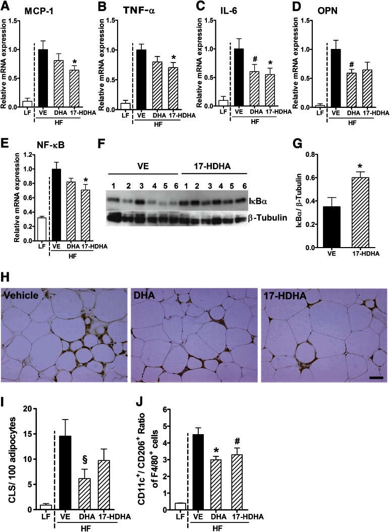
17-HDHA significantly decreases obesity-associated adipose tissue inflammation. HF-fed WT mice were treated with DHA, 17-HDHA, or vehicle (VE) control via intraperitoneal injection every 12 h for 8 days. 17-HDHA treatment reduced mRNA expression of the inflammatory genes for MCP-1 (Ccl2) (A), TNF-a (Tnf) (B), IL-6 (Il6) (C), OPN (Spp1) (D), and NF-κB (Nfkb1) (E) in gonadal adipose tissue of WT HF animals compared with VE-treated control group (n = 12–14 animals per group). (F) and (G) Immunoblot analysis and quantification of IκBα in gonadal adipose tissue of WT HF animals after VE and 17-HDHA treatment. The diagram shows means of the chemiluminescence intensity ratios from IκBα vs. β-tubulin, which was used as the loading control (G) (n = 6 animals per group). (H) Representative images of CLS formation in gonadal adipose tissue (scale bar = 50 µm). (I) Number of CLS counts per 100 adipocytes in gonadal adipose tissue after VE, DHA, or 17-HDHA treatment (n = 5–6 animals per group). (J) Flow cytometry analysis of the CD11c+CD206−-to-CD11c−CD206+ ratio of adipose tissue macrophages (F4/80+ cells) obtained from stromal vascular fractions after DHA and 17-HDHA treatment compared with VE control (n = 5–6 animals per group). For statistical analysis, DHA- and 17-HDHA–treated groups were compared with the VE-treated control group. All data are mean ± SEM. #P < 0.08; §P = 0.088; *P < 0.05. (A high-quality color representation of this figure is available in the online issue.)

