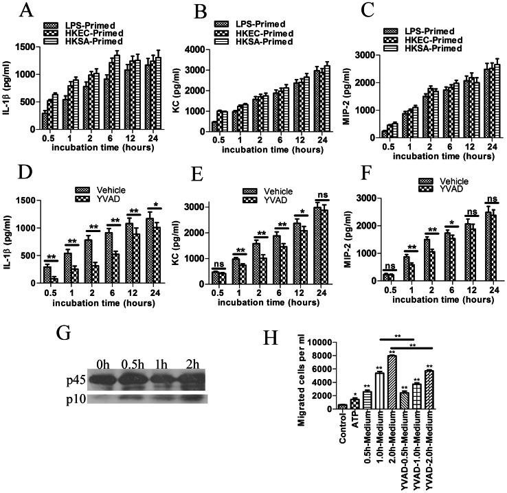
Figure 2. ATP stimulats cytokine and chemokine secretion and inflammasome activation directly and indirectly induces in vitro neutrophil chemotaxis.
(A, B and C) BMDMs were primed with LPS (500 ng/ml), heat killed E. coli 25922 (HKEC) (3×10⁸ CFU/ml) or heat killed S. aureus 25923 (HKSA) (3×10⁸ CFU/ml) and the medium was replaced with fresh serum-free medium and incubated with ATP (2 mM) for 0.5, 1, 2, 6, 12 and 24 hours. The levels of IL-1β, KC and MIP-2 were then assessed by ELISA. (D, E and F) To test the role of caspase-1 in the cytokine secretion, the cells were pre-incubated with the Caspase-1 inhibitor Ac-YVAD-cho (50 µM) for 30 min after LPS-priming, then incubated with ATP. The data are presented as the means±SEMs (n = 5). Values that are significantly different are indicated by asterisks; *(P<0.05), **(P<0.01). Values that showed no significant differences are marked “ns”. (G) BMDMs incubated with ATP (2 mM) for the indicated times were lysed and immunoblotted with Caspase-1 antibody (Santa Cruz). The appearance of the p10 subunit represented the activation of caspase-1 and the inflammasome. The presented graph is representative of at least three independent experiments. (H) ATP-stimulated culture medium was harvested, and the ability to induce neutrophil chemotaxis was assessed using 3-µm-pore transwells. The data are presented as the means±SEMs (n = 5). The number of migrated cells was compared with the control (containing fresh medium in the lower chamber), and the values that are significantly different are indicated by asterisks as follows: *(P<0.05), **(P<0.01).

