
Figure 2.
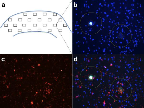
Plaque-centered stereology-based study. (a) Diagram represents a typical map of fields resulting from the random sampling of a paraffin section of temporal cortex using the parameters described in the Methods section. (b, c) Fields with Thioflavin-S (ThioS)-positive plaques present in the blue-green channel also exhibiting DAPI nuclear staining (b) were photographed and the corresponding images of glial cells in the red channel (c) were also taken. (d) Finally, the pairs of images from both channels corresponding to the same fields were merged in the ImageJ software, the sizes of the plaques were measured with the appropriate tool of the software, and numbers of glial cells with a DAPI-visible nucleus located within 50 μm from the edge of each plaque were manually counted.