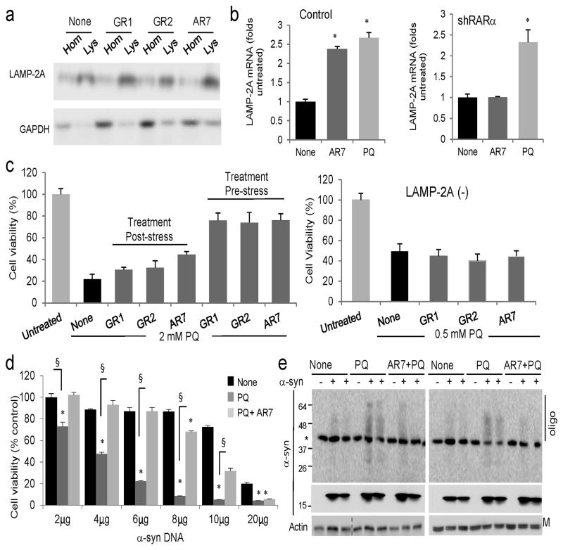
Figure 7. Effect of the retinoid derivatives in the cellular response against different stressors.
(a) Immunoblot for the indicated proteins in homogenates (Hom) and lysosomes (Lys) isolated from cells untreated (none) or treated for 12 h with 20μM of the indicated compounds. (b) mRNA levels of LAMP-2A in mouse fibroblasts control (Ctr) or knocked down (−) for RARα, and treated with AR7 or paraquat (PQ) as in a (n=4–5). (c) Cellular viability of control (left) or LAMP-2A(−) (right) fibroblasts exposed to 2 mM or 0.5 mM PQ, respectively, and treated with the indicated compounds for 12 h before or after the PQ treatment. (n=3). (d) Viability of mouse fibroblasts transfected with the indicated concentrations of a plasmid encoding α-synuclein and left untreated (none) or treated with 1 mM PQ alone or in the presence of 20μM AR7. (n=3). (e) Immunoblot for α-synuclein in the same cells as d. Top: higher exposure blot to highlight oligomeric (Oligo) species. *nonspecific band. M: monomer; All values are mean±S.E. Differences with cells untreated (*) or treated only with PQ (§) were significant for p<0.001. Full-length blots are shown in Supplementary Figure 21.

