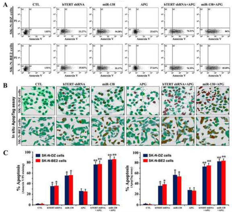
Fig. 3.
Flow cytometry and in situ ApopTag assay for detection and determination of amounts of apoptosis in SK-N-DZ and SK-N-BE2 cells. Treatments: control (CTL), hTERT shRNA plasmid (1 μg/ml) for 12 h, miR-138 mimic (50 nM) for 12 h, 100 μM APG for 24 h, hTERT shRNA plasmid (1 μg/ml) for 12 h + 100 μM APG for 24 h, and miR-138 mimic (50 nM) for 12 h + 100 μM APG for 24 h. (A) Annexin V-FITC/PI double staining followed by flow cytometry to detect the early phase of apoptosis. (B) In situ ApopTag assay to detect the late phase of apoptosis. (C) The percentages of apoptotic cells are shown in bar diagrams based on results from three independent experiments. Difference between untreated CTL and a treatment group was considered significant at *p < 0.05 or **p < 0.01.

