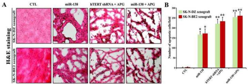
Fig. 6.
Evaluation of histopathological changes in the SK-N-DZ and SK-N-BE2 tumors after treatments. Treatments: control (CTL, no treatment for 21 days), tumor bearing mice (7 days after tumor implantation) were injected at the tumor site with miR-138 mimic (50 μg DNA/injection/mouse), hTERT shRNA plasmid (50 μg DNA/injection/mouse) + APG (10 μg/injection/mouse), and miR-138 mimic (50 μg DNA/injection/mouse) + APG (10 μg/injection/mouse) on alternate days for 2 weeks. All animals were then sacrificed for analyzing the histopathological changes in the tumors. (A) H&E staining to examine histopathological changes in the xenograft sections. (B) The apoptotic cells in SK-N-DZ and SK-N-BE2 xenografts were shown in bar diagrams based on observation from three independent microscopic fields. Difference between CTL group and a treatment group was considered significant at *p < 0.05 or **p < 0.01.

