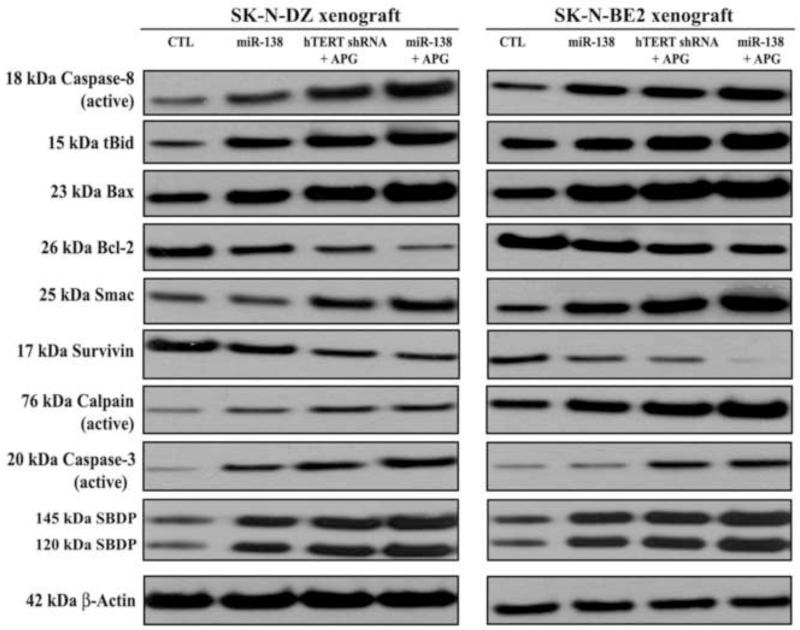
Fig. 7.
Changes in expression of apoptotic proteins in SK-N-DZ and SK-N-BE2 tumors after treatments. Treatments: control (CTL, no treatment for 21 days), tumor bearing mice (7 days after tumor implantation) were injected at the tumor site with miR-138 mimic (50 μg DNA/injection/mouse), hTERT shRNA plasmid (50 μg DNA/injection/mouse) + APG (10 μg/injection/mouse), and miR-138 mimic (50 μg DNA/injection/mouse) + APG (10 μg/injection/mouse) on alternate days for 2 weeks. All animals were then sacrificed for analyzing the molecular changes in the tumors. Representative Western blots (n ≥ 3) to show expression and activation of selective molecules involved in the induction of extrinsic and intrinsic pathways of apoptosis. Expression of β-actin was used as a loading control.

