
Fig. 3.
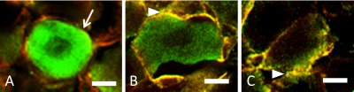
NGF-IR at satellite glial cells (SGCs) or neurons in the trigeminal ganglion (TG). Three types of NGF-IR were observed: (A) the neuron was immunopositive for NGF (arrow), but surrounding SGCs were negative [N(+)S(–)]; (B) both the neuron and surrounding SGCs (arrowhead) were positive [N(+)S(+)]; and (C) the SGCs were positive, but the neuron they surrounded was negative [N(–)S(+)]. Green; NGF-IR cells, red; GS-immunoreactive cells, and yellow; NGF and GS double positive cells. Bars=10 µm (A, B, and C).