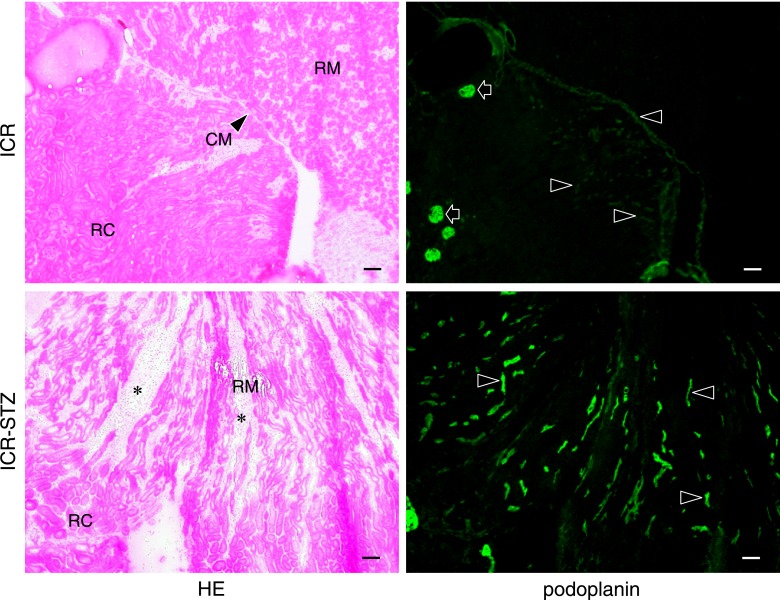
Fig. 3.
Distribution of lymphatic vessels in the ICR and ICR-STZ mouse kidneys. The immunostained sections were re-stained by HE staining. The HE staining shows that the kidney tissue is collapsed by edema (asterisks): renal tubules and the peritubular space expanded in the renal medulla (RM) in the ICR-STZ mice compared with the ICR control mice. In the ICR control mice, podoplanin-stained glomeruli (arrows) are present in the renal cortex (RC) near the renal surface and podoplanin-stained lymphatic vessels (arrowheads) are present in the cortex near the cortico-medullary border (CM), but absent in the renal medulla (RM). In the ICR-STZ mice, many podoplanin-stained lymphatic vessels (arrowheads) are present in the cortex and medulla at the expanded peritubular space (asterisks). Bar=100 µm.

