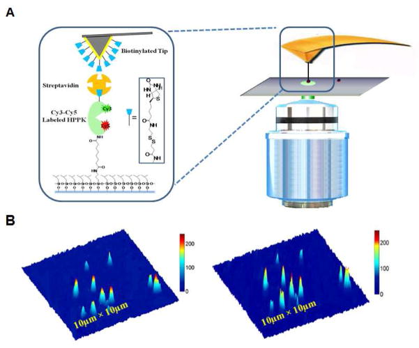
Figure 2.
(A) Single-molecule AFM-FRET ultra nanoscopy, the zoomed panel in the left presents schematic diagram of one FRET dye-pair (donor-acceptor: Cy3–Cy5) labeled HPPK molecule tethered between a glass cover-slip surface and a handle (biotin group plus streptavidin), and another biotin group is modified on AFM tip. (B) Single-molecule fluorescence photon counting images of the donor (Cy3, left) and accepter (Cy5, right). Each feature is from a single HPPK enzyme labeled with Cy3–Cy5 FRET dyes.

