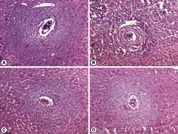
Fig. 2.
Photomicrograph of liver granulomas of S. mansoni-infected vector-vaccinated control and infected SMALDO vaccinated groups. (A) Vaccinated control group: a large granuloma formed of a central egg with living miracidium surrounded by a large number of eosinophils, neutrophils, and lymphocytes. (B) IP, (C) SC, and (D) IM groups showing small cellular granulomas, formed of an egg in the center surrounded by lymphocytes and few eosinophils and thin collagen fibers (H-E stain, ×200).

