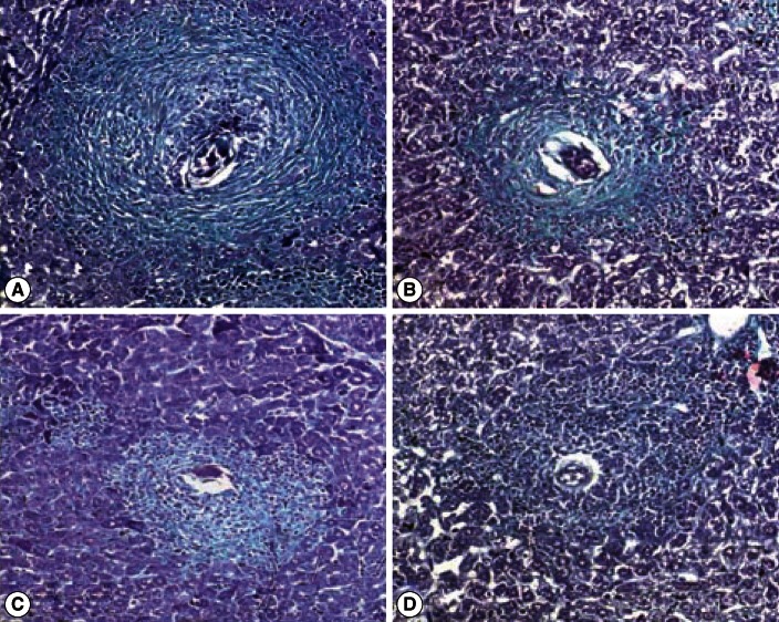
Fig. 3.
Photomicrograph of liver granulomas of S. mansoni-infected vector-vaccinated control and infected SMALDO vaccinated groups.(A) Vaccinated control group: a large fibrocellular granuloma formed of a central egg surrounded by inflammatory cells and deposited collagen fibers. (B) IP, (C) SC, and (D) IM groups showing small cellular granulomas formed of a central egg, surrounded by lymphocytes, histiocytes, fibroblasts, and thin concentric collagen fibers (Masson's trichrome stain, ×200).

