
Figure 2.
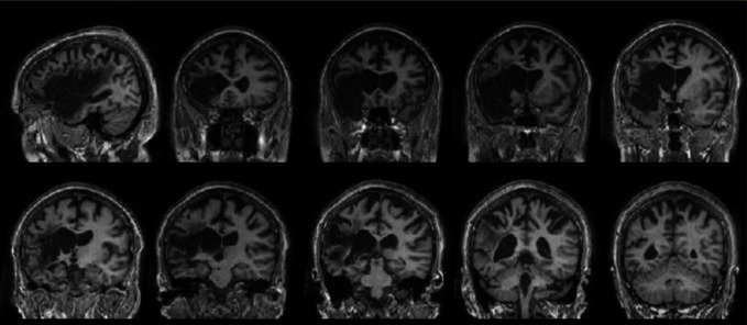
MRI of SKO acquired 9 years post-stroke, demonstrating an extensive left fronto-temporo-parietal lesion. Presented are a single sagittal slice, with nine coronal slices from anterior to posterior through the lesion area.
Official websites use .gov
A
.gov website belongs to an official
government organization in the United States.
Secure .gov websites use HTTPS
A lock (
) or https:// means you've safely
connected to the .gov website. Share sensitive
information only on official, secure websites.
MRI of SKO acquired 9 years post-stroke, demonstrating an extensive left fronto-temporo-parietal lesion. Presented are a single sagittal slice, with nine coronal slices from anterior to posterior through the lesion area.