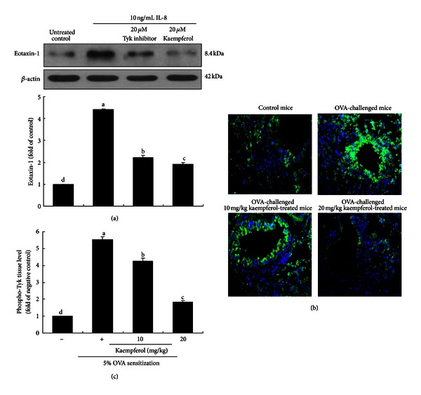
Figure 5.

Effect of Tyk2 inhibition on eotaxin-1 expression in IL-8-stimulated BEAS-2B cells (a). BEAS-2B cells were treated with 20 μM Tyk inhibitor and 20 μM kaempferol exposed to 10 ng/mL IL-8 for 8 h. Cell extracts were subjected to western blot analysis with a primary antibody against eotaxin-1 (3 separate experiments, (a)). β-actin was used as an internal control. The bar graphs (mean ± SEM) in the bottom panel represent densitometric results. Immunofluorescence analysis showing inhibition of Tyk2 activation in OVA-challenged mouse lung tissues by kaempferol (b and c). Cytoplasmic Tyk2 was visualized with an FITC-conjugated secondary antibody. Nuclear staining was done with DAPI. Tyk2 was identified as green staining and quantified by using an optical microscope system (c). Each photograph is representative of four mice. Magnification: 200-fold. Means without a common letter differ, P < 0.05.
