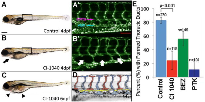
Figure 2. Formation of Thoracic Duct in Treated and Untreated Zebrafish Embryos.
Embryos treated with MAPK (Mek1/2) pathway inhibitor CI-1040 developed progressive pericardial (black arrow) and diffuse (black arrowhead) edema over time (panels A-C). Microscopic magnification of boxed areas in A and B are noted. TD development was evaluated over a length of 6 ISVs and located between the dorsal aorta and posterior cardinal vein (labeled) (panel A′). Panel D shows a schematic indicating the thoracic duct in yellow. In fish treated with CI-1040, loss of TD formation and lack of presumed LECs (white arrows) were noted (panel B′). Compared with controls, all treatment groups exhibited statistical prevention of formation of TD (p<0.0001, Chi squared test). In addition, CI-1040 more strongly prevented formation compared with BEZ (p<0.0001, OR=3.9) Error bars indicate standard deviation between experiments (panel E). Scale bar A 300 μm; A′ 100 μm.

