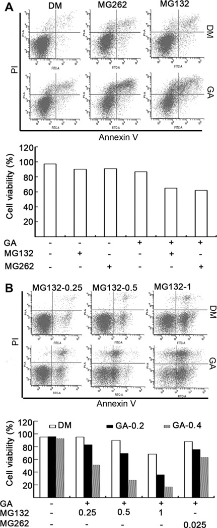
Fig. 2.
GA sensitizes malignant cells to the proteasome inhibitor-induced cell death. (A) H22 cells were treated with MG132 (0.5 µM) or MG262 (0.025 µM) in the presence or absence of GA (0.4 µM) for 24 h. The treated cells were collected and stained with FITC Annexin V and propidium iodide (PI), followed by flow cytometry analysis. The results of flow images (the upper panel) and summarized bar graphs (the lower panel) were presented. (B) K562 cells were treated with MG132 (0.25, 0.5, 1 µM) or MG262 (0.025 µM) in the presence or absence of GA (0.2, 0.4 µM) for 24 h, followed by staining with Annexin V and PI, and flow cytometry analysis.

