
Fig. 3.
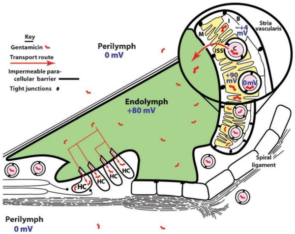
Schematic diagram of the cochlear duct cytoarchitecture and its electrophysiological environments. The stria vascularis, lining the spiral ligament on the inside lateral wall of the bony cochlear shell, contains basal (B) and marginal (M) cells connected together by tight junctions that form an impermeable paracellular barrier to solutes. Circulating aminoglycosides within strial capillaries (C) are preferentially transported through the strial blood–labyrinth barrier consisting of tight junction-coupled endothelial cells, into the intra-strial space (ISS). From there, aminoglycosides are trafficked through marginal cells into endolymph, and enter hair cells (HC) across their apical surfaces by endocytosis and non-selective cation channel permeation. The electrical potentials of various fluid compartments, separated by tight junction-coupled endothelial and epithelial cell barrier layers, are also indicated. Endolymph has a +80 mV, and hair cells have a resting potential of −60 to −75 mV, generating a considerable electrophoretic driving force across the apical endolymphatic membranes of hair cells.