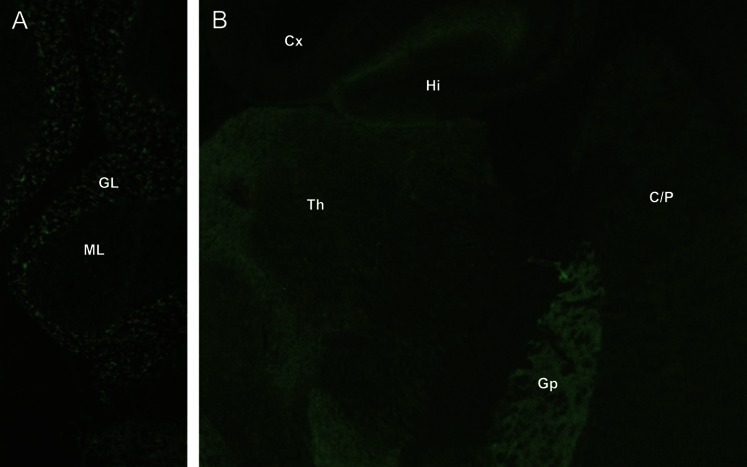
Figure. GAD65-IgG detected by indirect immunofluorescence on mouse cerebellum.
(A) Cerebellum, (B) other brain regions (coronal section). Characteristic synaptic staining pattern is most prominent in the granular layer (GL) of cerebellum and globus pallidus (Gp). Less intensely stained regions include molecular layer of cerebellum (ML), thalamus (Th), caudate and putamen (C/P), hippocampus (Hi), and cerebral cortex (Cx). GAD65 = glutamic acid decarboxylase 65-kDa isoform; Ig = immunoglobulin.

