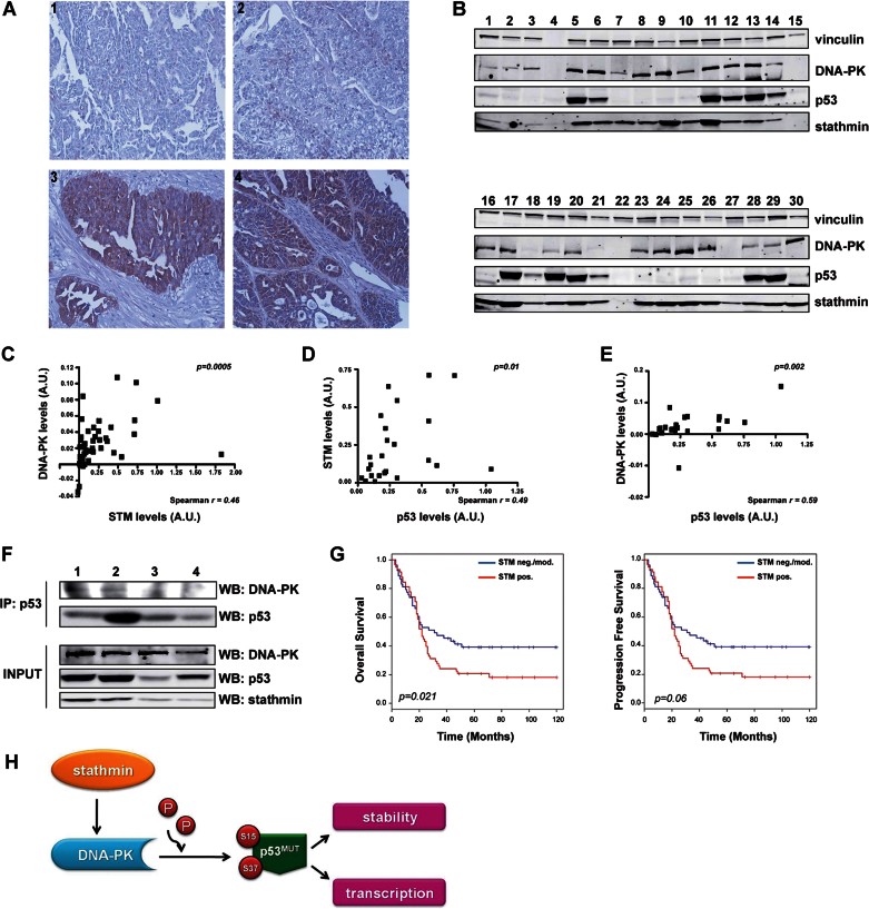
Figure 8.
Stathmin overexpression identifies low-surviving ovarian cancer patients.
Source data is available for this figure in the Supporting Information.
A. Representative immunohistochemical staining of formalin-fixed, paraffin-embedded EOC tissue sections negative/moderate (panel 1: serous G3; panel 2: endometrioid G2) and high (panel 3: serous G2; panel 4: serous G3) for stathmin expression. Original magnification 200×.
B. Representative Western blot analyses of p53, stathmin and DNA-PK expression in total protein extracts from primary high-grade serous carcinoma samples.
C–E. Spearman's correlation analysis between stathmin and DNA-PK (C), p53 and stathmin (D) or p53 and DNA-PK (E) expression in primary serous carcinomas.
F. Co-immunoprecipitation of p53 and DNA-PK in primary high-grade serous carcinoma samples. Total protein lysates from tumours expressing high (lanes 1, 2) or low (lanes 3,4) levels of stathmin expression (input, lower panels) were immunoprecipitated using anti-p53 antibody and then immunoblotted using anti-DNA-PK and anti-p53 antibodies, as indicated.
G. Kaplan Meyer estimate of overall (left) and progression free (right) survival, following stratification for stathmin expression. p values were calculated using the log-rank test.
H. Schematic representation depicting the role of stathmin in EOC carrying a p53MUT protein. High stathmin levels in EOC are necessary for proper expression of DNA-PK that, in turn, positively regulates p53MUT stability and transcriptional activity through S15/S37 phosphorylation.

