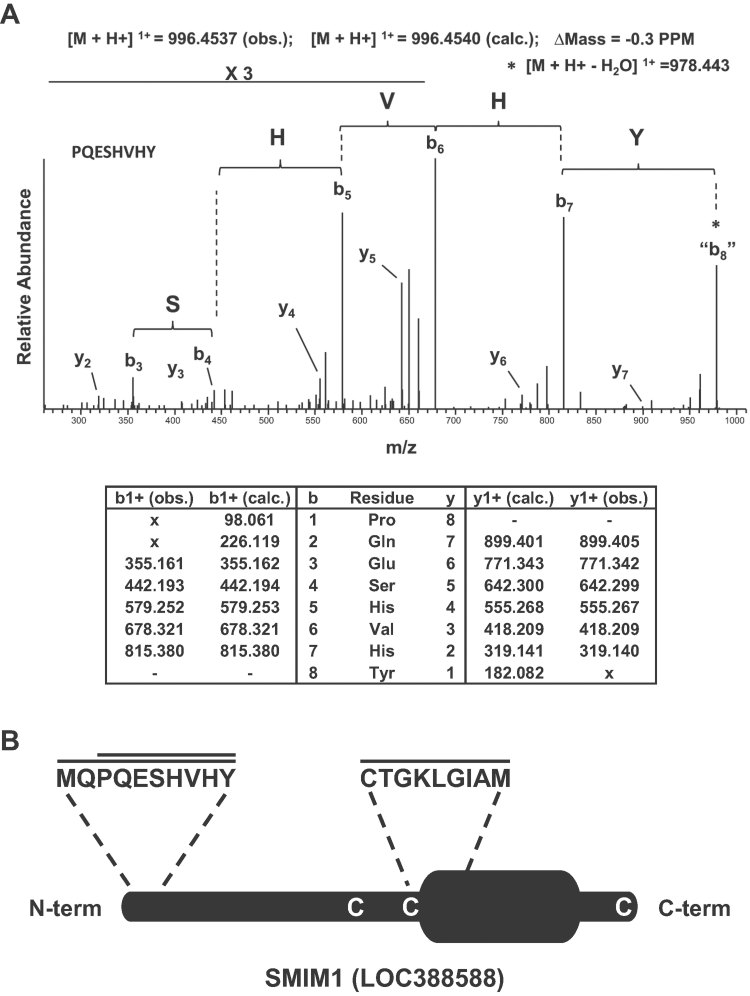
Figure 3.
Mass spectrometry-based identification of the Vel antigen carrier as SMIM1.
A. The top panel shows the high resolution MS/MS spectrum acquired in the orbitrap mass spectrometer corresponding to the SMIM1 peptide PQESHVHY that was annotated following de novo peptide sequencing as described in the Methods Section. The bottom panel provides the corresponding calculated (calc.) and observed (obs.) m/z values of the singly charged y- and b-type ions.
B. Schematic representation of the SMIM1 protein showing the predicted transmembrane domain, the peptides that were identified by mass spectrometry, and the three cysteine residues potentially involved in dimer formation.

