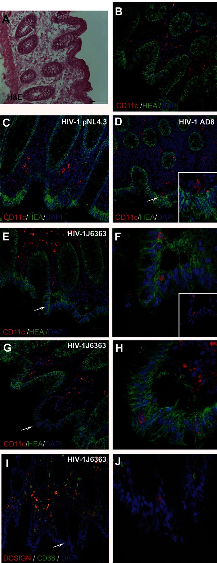
Figure 4.
R5 HIV-1 induces migration of DCs through the colonic epithelium.
Colonic tissue was either left untreated (A and B) or incubated with X4 HIV-1pNL4.3 (C), R5 HIV-1AD8 (D) or R5 HIV-1J6363 (E–J) (at 50 ng of p24 Ag) for 30 min. CD11c+ cells were detected in the colonic lamina propria of untreated (B) and HIV-1pNL4.3 treated (C) tissues but not in between epithelial cells. Following R5 HIV-1 incubation protruded dendrites (D) or whole DCs (E–H) were observed inside the epithelium. Moreover DCs (DCSIGN+/CD68− cells) but not Mϕ (DCSIGN+/CD68+ and DCSIGN-CD68+ cells) migrated through the epithelium upon HIV-1 stimulation (I and J). Cryosections were fixed with 4% PFA and immunostained with Hematoxylin–eosin (A), or for human epithelial antigen (mouse anti-HEA-FITC, green), DAPI (nuclei; blue) and either mouse anti-human CD11c + Alexafluor594 goat anti-mouse IgG (DCs; red) (B–H) or mouse anti-human DC-SIGN-PE (DCs and Mϕ; red) and mouse anti-human CD68+ Alexafluor488 goat anti-mouse IgG1k (monocytes/Mϕ; green) (I and J). The inset in panel (D) and (F) is magnified 3×. Panels (F), (H) and (J) are magnification (zoom 3×) of the area indicated by arrow in (E), (G) and (I), respectively. The inset of panel (F) evidences DCs after hiding of HEA channel. Scale bar: 50 µm in B–E, G and I. Each Figure is representative of results from five donor tissues.

