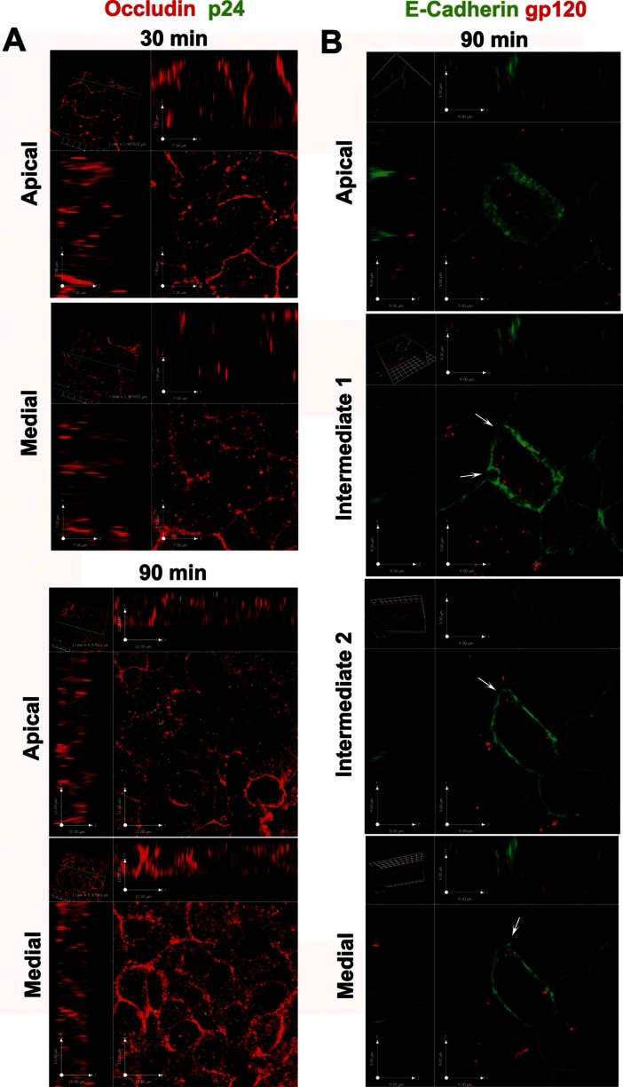
Figure 9.
HIV-1 penetrates within and in between epithelial cells.
A. Virions (stained with mouse anti-p24 + Alexafluor488 goat anti-mouse IgG, green) were mainly localized at the apical surface of Caco-2 cells at 30 min but also inside the cytoplasm at 90 min of incubation with R5 HIV-1AD8 (20 ng of p24 Ag). Shown are CM single plane cross sectional images of a Caco-2 monolayer (rabbit anti-human Occludin + Alexafluor594 goat anti-rabbit IgG; red) taken at apical and medial level of the cell layer.
B. R5 HIV-1AD8 (red; visualized with human anti-gp120 monoclonal antibody 2G12 + Alexafluor594 goat anti-human IgG) localized inside the cytoplasm of epithelial cells and in intrajunctional spaces (indicated by arrows). Shown are four single plane cross sectional images, taken from the apical to the medial plane along the z-axis of the Caco-2 monolayer (mouse anti-human E-Cadherin + Alexafluor488 rabbit anti-mouse IgG2a; green) incubated with R5 HIV-1J6363 (at 5 ng of p24 Ag) for 90 min. Results are of one representative experiment out of three.

