
Figure 2. Histologic features.
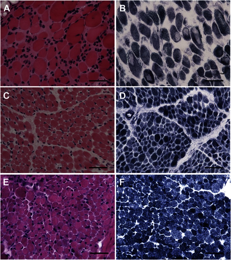
(A) Skeletal muscle of patient 6 demonstrates a large majority of rounded fibers with severe variation in size and occasional fibers with internally placed nuclei. (C) Patient 1 demonstrates mild increase in connective tissue and significant variation in fiber size, but no internally placed nuclei. (E) Patient 5, similarly to patient 1, demonstrates variation in fiber size and shape, but not significant fibrosis; in addition, scattered fibers have internally placed nuclei. Whereas oxidative enzyme stain (NADH-TR) reveals no cores in patients 1 (D) and 5 (F), there are numerous, well-defined cores centrally and peripherally located present in patient 6 (B). A, C, E: hematoxylin & eosin; B, D, F: NADH-TR; A–F calibration bar 50 µm. NADH-TR = nicotinamide adenine dinucleotide dehydrogenase–tetrazolium reductase.