
Figure 3. Histologic features.
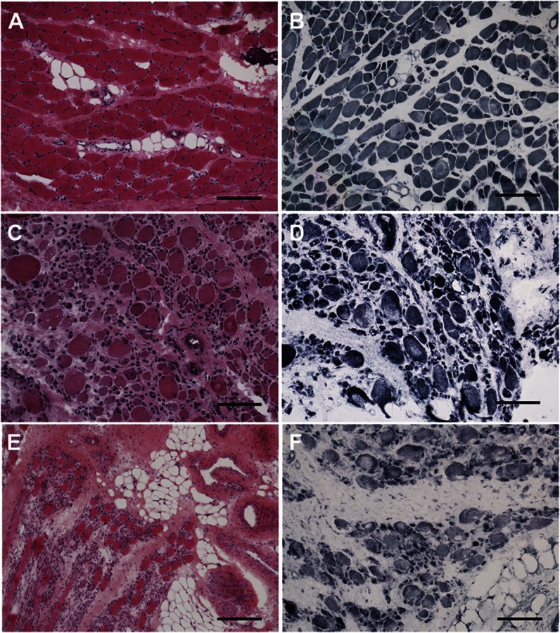
(A) Mild fibrosis and adipose tissue infiltration with moderate variation in size and shape but scattered fibers with internally placed nuclei are seen in patient 4. (C) Patient 2 demonstrates rounded fibers, most of which are surrounded by excess connective tissue, occasional dark, hypercontracted-appearing fibers, and severe variation in size with clusters of small fibers. (E) In patient 3, there is pronounced increase in adipose and connective tissue with numerous minute fibers and rounded hypertrophic fibers. Oxidative enzyme stain (NADH-TR) reveals scattered fibers with ill-defined cores in patients 2 (D) and 3 (F) and unevenness of stain with ill-defined cores in patient 4 (B). A, C, E: hematoxylin & eosin; B, D, F: NADH-TR; C, D, F calibration bar 100 μm; E, A, B calibration bar 200 μm. NADH-TR = nicotinamide adenine dinucleotide dehydrogenase–tetrazolium reductase.