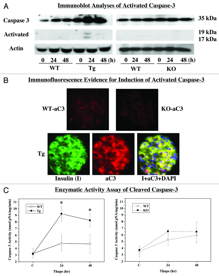
Figure 4. ER stress-induced activation of caspase-3 in WT, RIP-iPLA2β-Tg and iPLA2β-KO islets. Islets (200/condition) from WT, iPLA2β-Tg and iPLA2β-KO mice were cultured O/N at 37°C under an atmosphere of 5%CO2/95% air and then treated with either vehicle (DMSO) or with thapsigargin (Thaps, 2 μM) for up to 48 h and assessed for expression of cleaved (i.e., activated) caspase-3. (A) Immunoblotting analyses. Total and cleaved caspase-3 (ac3) expressions were determined using 30 μg aliquots of islet lysate. (B) aC3 Immunofluorescence. Representative paraffin islet sections (8–10 μm) co-stained for nuclei (DAPI), insulin (I) and aC3. (C) aC3 activity assay. Islet lysates were prepared and activity in 30 μg aliquot of protein was assayed using a colorimetric-based protocol. (*Significantly different from WT group, p < 0.05.)

An official website of the United States government
Here's how you know
Official websites use .gov
A
.gov website belongs to an official
government organization in the United States.
Secure .gov websites use HTTPS
A lock (
) or https:// means you've safely
connected to the .gov website. Share sensitive
information only on official, secure websites.
