
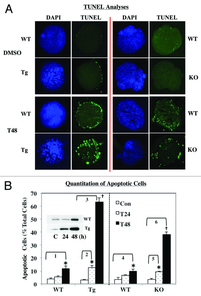
Figure 7. ER stress-induced apoptosis in WT, RIP-iPLA2β-Tg and iPLA2β-KO islets. Islets (200/condition) from WT, iPLA2β-Tg and iPLA2β-KO mice were cultured O/N at 37°C under an atmosphere of 5%CO2/95% air and then treated with either vehicle (Con, DMSO) or with thapsigargin (T, 2 μM) for up to 48 h. (A) TUNEL analyses. Intact islets were processed for TUNEL analyses to visualize cells undergoing apoptosis, as reflected by punctate green fluorescence. (B) Quantitation of apoptotic cell number. To assess the incidence of apoptosis, islets were dispersed into individual cells and flow cytometry protocol was used to determine the number of apoptotic cells, relative to total cell number in each preparation. The means ± SEM of the percentage of apoptotic cells in each group are presented. (*Significantly different from corresponding WT group and †significantly different from corresponding T24 and WT groups, p < 0.05, n = 3–5). Fold increases between groups are as follows: 1, 3.09 ± 0.50; 2, 4.50 ± 0.54; 3, 22.24 ± 1.18; 4, 2.73 ± 0.52; 5, 2.70 ± 0.19; and 6, 10.96 ± 0.64). Inset. Induction of iPLA2β during ER stress. Cytosol (30 μg WT and 10 μg iPLA2β-Tg) from thapsigargin-treated islets was resolved by SDS-PAGE, protein transferred to immunoblots and probed for iPLA2β.