Fig. 10.
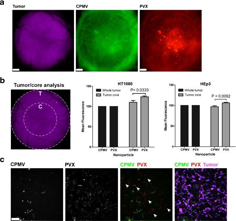
Intravital imaging of VNP uptake in human tumor xenografts in the CAM. a Avian embryos bearing vascularized GFP-expressing human fibrosarcoma HT1080 or human epithelial carcinoma HEp3 tumors (magenta) were co-injected with 515 × 13 nm filamentous PVX-PEG-A555 (red) and 30 nm-sized icosahedron CPMV-PEG-647 (green) and visualized 4 h after injection. Scale bar = 190 μm. b The analysis of whole tumor uptake of CPMV and PVX nanoparticles compared to uptake only in the tumor core was assessed using distinct ROIs (left panel) in HT1080 (middle panel) and HEp3 (right panel) tumors. While whole tumor localization of CPMV and PVX were comparable, PVX accumulated in the core of tumors to a significantly higher degree than CPMV (unpaired t test). c The localization of nanoparticles was assessed in 8 μ sections of the tumor core using fluorescence microscopy. CPMV (green) and PVX (red) are visualized in the tumor (magenta). While CPMV was visualized in punctate foci, PVX was distributed throughout the tumor in areas devoid of CPMV (white arrowheads). Reproduced with permission from Shukla et al., Mol. Pharm. 2013 [95]

