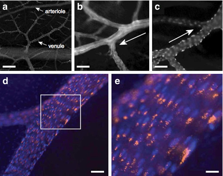
Fig. 8.
Intravital fluorescence imaging of chick CAM vasculature and location of A555-labeled, fluorescent CPMV. a Fluorescence imaging looking down through surface chick CAM showing multiple levels of vasculature; scale bar is 100 μ. b CAM arteriole. Scale bar is 22 μ. c CAM venule; scale bar is 22 μ. Arrows in b and c denote blood flow direction. d, e Intravital image of large CAM vein, CPMV-A555 in orange, endothelial cells nuclei in blue. CPMV particles are restricted in the perinuclear compartment in vascular endothelial cells. Scale bar is 16 μ in d and 5.5. μ in e. Reproduced with permission from Lewis et al., Nature Medicine 2006 [95]

