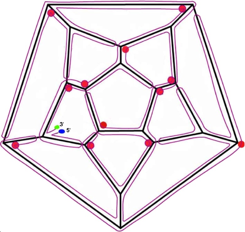
Fig. 2.
Organization of a hypothetical secondary structure for PaV RNA, in the stalactite conformation [9]. This is just one solution for mapping the RNA onto the dodecahedral cage in a fashion that is consistent with the data from X-ray crystallography and cryo-electron microscopy. The 5′ and 3′ ends are indicated. Each edge of the dodecahedral cage contains an antiparallel RNA double helix. Red circles indicate three- and four-way junctions at those vertices where RNA stalactites drop down into the interior of the virus to connect with that part of the RNA not on the dodecahedral cage

